Kidney Stones

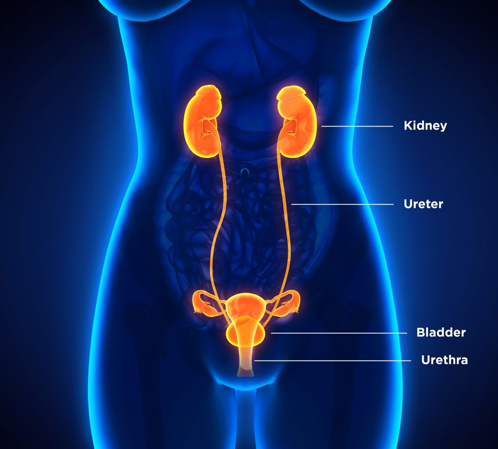
Female Urinary System

HOW AND WHERE KIDNEY STONES OCCUR

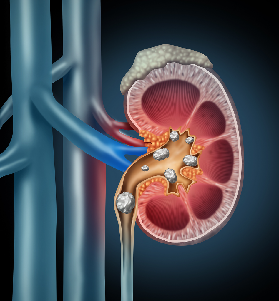
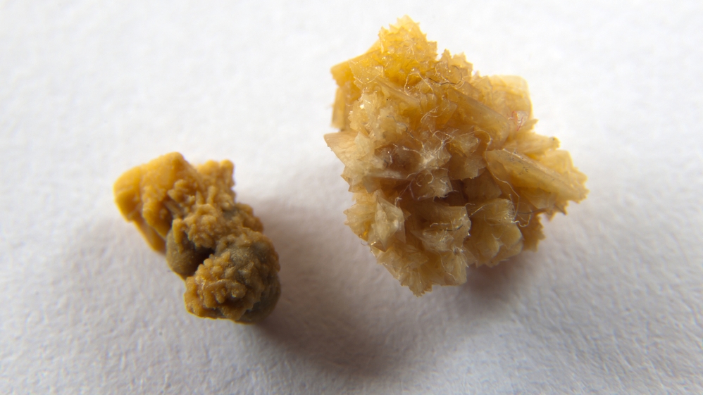
Most urinary stones (urolithiasis in medical terms) arise as a result of a change in the natural balance of minerals and water in the urine. They consist of salt and minerals in the urine, which stick together and form small clots. The stones are rarely larger than a grain of rice, but they can grow to several centimeters in diameter and in some cases they even fill the entire renal collecting system.

They can stay in the kidneys or exit the body through the urinary tract. The urinary tract is the system that generates and excretes urine. It comprises the kidneys, the ureters (which connect the kidneys to the bladder), the bladder and the urethra, through which urine is transported from the bladder out of the body. Depending on the diagnosed condition, a distinction is made between kidney, urinary and bladder stones. Doctors distinguish between the type of salt that the stone comprises, for example:

  • Calcium oxalate stones (70-75%) [1, 2]
  • Uric acid stones (up to 10%) [1, 2]
  • Infectious stones, e.g. “Struvite stones” consisting of magnesium ammonium phosphate (approx. 10-15%) [1-3]

Factors that can change the balance of substances in the urine and are therefore a common cause of kidney and urinary stones include the following:

  • Insufficient water intake, which can result in salts and minerals sticking together to form small stones. [2, 4]
  • A protein- or sodium-rich diet [5, 6]
  • Frequent urinary tract infections [2, 4]
  • Metabolic syndrome, obesity, diabetes and gout [1, 7-10]
  • A regulatory disorder of the parathyroid glands (hyperparathyroidism) [1, 2, 4]
  • Bowel surgery, gastric bypass and chronic bowel disease (Crohn’s disease) [1, 2, 4]
Delta III Coupling

TREATING KIDNEY STONES USING ESWL

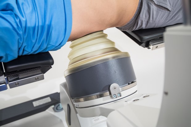
First introduced in 1980, extracorporeal shock wave lithotripsy (ESWL) is now a standard treatment option for dealing with kidney and urinary stones. Shock wave lithotripsy is a gentle way of removing urinary stones as it does not require surgery. Most stones can be treated successfully using this method. It involves sending shock waves into the body from the outside. These waves travel through the tissue and meet at a focal point on the stone, causing it to shatter. If this treatment is successful, the fragments of stone then exit the body naturally with the urine.

The shock waves work through the skin, so there is no need for any invasive intervention such as surgery. ESWL treatment is usually not very painful. However, if patients do feel any pain, the doctor treating them can inject a painkiller directly into a vein to ensure that it takes effect quickly. Shock wave lithotripsy takes approximately 30 to 60 minutes and can usually be performed as an outpatient procedure. Using state-of-the-art equipment, all stone deposits along the urinary tract can be treated while the patient is lying comfortably on their back. With the help of ultrasound and X-ray imaging, the shock wave generator is positioned so that the shock waves are concentrated on the stone. X-rays and/or ultrasounds are also used to check the success of the treatment during and after the procedure. [11]

Depending on the size and position of the stones, high success rates can be achieved with just one round of treatment, which often can be done out patient. Scientific studies indicate that stones are completely cleared in up to 90% of well-selected patients who undergo ESWL and in more than 70% of cases only one treatment session is required. [12, 13]

URS Procedure

Endoscopic Removal of Kidney or Urinary Stones (URS)

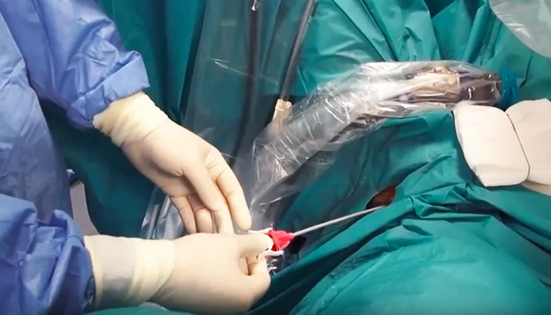
Larger kidney stones are often surgically removed. Endoscopic removal of kidney or urinary stones is usually carried out under general anesthetic with the patient in the lithotomy position (on their back with legs spread apart). With the help of an endoscope, fine instruments are inserted through the urethra and bladder and up into the ureter until they reach the stone. The stone is then broken up, either mechanically or by laser, so that the pieces can be excreted or removed endoscopically using small forceps or baskets. The technical term for this endoscopic treatment is ureterorenoscopy (URS), or retrograde intrarenal surgery (RIRS) if the stone is located in the kidney. Usually the patient can leave the hospital after a recovery time of two days. [11]

At the end of this endoscopic operation, a ureteral stent (double-J catheter) is usually inserted under general anesthetic to prevent any obstruction of the urine flow and to avoid putting any strain on the patient. The double-J catheter is removed again in an outpatient procedure after one to two weeks. [11]

Videos:

http://www.maestro-portal.eu/procedure/detail/1 (Dornier laser)

http://www.maestro-portal.eu/procedure/ (Dornier laser)

PCNL Treatment

Surgical Kidney Stone Removal Through the Skin (PCNL)

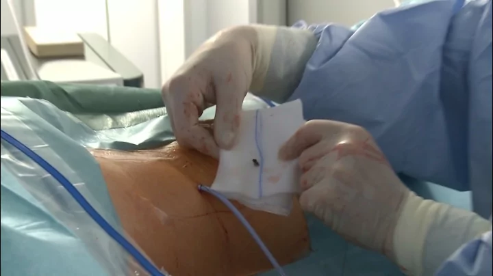
This procedure is usually only carried out in the case of large and complex kidney stones, such as staghorn calculi. A staghorn calculus is a stone that fills large sections of the renal pelvis or one or more of the renal calyces. Percutaneous nephrolitholapaxy (PCNL) involves removing kidney stones by creating an artificial opening in the back. To do this, the kidney is punctured from the flank directly through the skin. This allows an endoscope to be inserted into the renal collecting system, where the stone is located. This operation is generally performed under general anesthetic. The urinary tract is prepared for the operation with the patient in the lithotomy position. The patient is then moved onto their stomach so that they are lying face down. This allows the surgery itself to be performed on the back.

The puncture procedure is carried out under visual control using ultrasound and X-ray imaging. Once the endoscope – which can be as thick as a pencil – has been inserted into the kidney, the stone can be broken up with an ultrasonic or laser probe and the pieces can then be removed. To finish off the operation, a catheter is inserted into the kidney (urinary diversion outwards via the flank) or a ureteral stent (internal diversion) is put in to prevent any obstruction of the urine flow.

The patient usually has to stay in hospital for three to five days. If the procedure is performed by an experienced surgeon, the complication rate is usually low. [11]

Recommended Stone Treatment ENG

Which Kidney Stone Treatment Method is Right for Me?

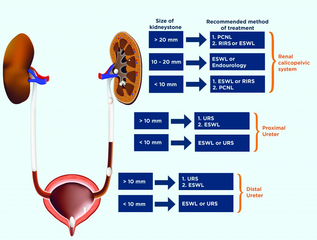
Kidney stones are treated using either extracorporeal shock wave lithotripsy (ESWL) or a surgical procedure such as URS or PCNL. The relevant medical associations (e.g. the German Society of Urology or the European Association of Urology) make recommendations on how to choose the best treatment method. These medical association recommendations are made available to urologists in the form of guidelines. They are based on the latest scientific knowledge obtained from medical studies. As of October 2017, the guidelines issued by the European Association of Urology (EAU)[1] on treating kidney and urinary stones are as follows:

The key factors are the location and size of the stone (first box), which are estimated first of all through diagnostic investigation. The second box shows the recommended treatment methods, which are listed either in order of preference (1 and 2) or as equal options. If, for example, you have an 8 mm stone in the proximal ureter (in a section of the ureter close to the bladder), the EAU recommends treatment with ESWL or URS.

Kidney Stone Reveal

Which Kidney Stone Treatment Method is Right for Me? – Part 2

Although endourological (URS) stone therapy usually involves general anesthesia and a hospital stay and can therefore be associated with certain risks for the patient, more and more urologists are showing a preference for this method. One reason frequently given for this is that they enable the stone to be removed completely and immediately with just one operation. The need for secondary treatment is often cited as a disadvantage of extracorporeal shock wave lithotripsy. As already mentioned, however, ESWL yields high success rates (up to >90% in well-selected patients/stones), in many cases with only one round of treatment needed. This non-invasive treatment method also offers some important advantages: shock wave treatment does not harbor the risks posed by invasive methods with regard to anesthesia, infection, surgery and hospitalization, and is often carried out on an outpatient basis. If secondary treatment is required, ESWL therapy is usually, in the same respects, less stressful for patients than the invasive URS and PCNL procedures described. [14, 15]

Your urologist will take various parameters into account when deciding on the right treatment for your kidney or urinary stone. In addition to the location and size of the stone, these may include the stone’s chemical composition, any pre-existing conditions and the anatomical characteristics of your body.

Non-invasive treatment such as ESWL has clear advantages. Your doctor will certainly be happy to arrange a consultation with you to explain whether lithotripsy is a viable option in your case and to tell you about possible side effects and complications, such as renal colic or haematoma.

Before the first lithotripsy (kidney stone disintegration) procedure was carried out on humans in 1980, patients had no choice but to undergo open surgery. Nowadays, more than 500,000 patients worldwide are treated using ESWL each year. This treatment offers a simple way of breaking up suitable stones. Dornier MedTech, the inventor of extracorporeal shock wave lithotripsy (ESWL), boasts 30 years of experience in this area and hundreds of its ESWL devices are in use around the world.

Kidney Stone Example

References

  1. C. Türk (Chair), A.N., A. Petrik, C. Seitz, A. Skolarikos, A. Tepeler, K. Thomas Guidelines Associates: S. Dabestani, T. Drake, N. Grivas, Y. Ruhayel. European Urolithiasis Guidelines. 2017 [cited 2017 27 September]; Available from: https://uroweb.org/guideline/urolithiasis/.
  2. EAU Patient Information. [cited 2018 January 9]; Available from: http://patients.uroweb.org/wp-content/uploads/sites/2/06_Basic-info.pdf.
  3. Flannigan, R., et al., Renal struvite stones–pathogenesis, microbiology, and management strategies. Nat Rev Urol, 2014. 11(6): p. 333-41.
  4. EAU Patient Information – Causes of kidney and ureteral stones. [cited 2018 January 9]; Available from: http://patients.uroweb.org/wp-content/uploads/sites/2/01_Causes-of-kidney-and-ureteral-stones.pdf.
  5. Breslau, N.A., et al., Relationship of animal protein-rich diet to kidney stone formation and calcium metabolism. J Clin Endocrinol Metab, 1988. 66(1): p. 140-6.
  6. Kok, D.J., et al., The effects of dietary excesses in animal protein and in sodium on the composition and the crystallization kinetics of calcium oxalate monohydrate in urines of healthy men. J Clin Endocrinol Metab, 1990. 71(4): p. 861-7.
  7. Ferraro, P.M., et al., Dietary and Lifestyle Risk Factors Associated with Incident Kidney Stones in Men and Women. J Urol, 2017. 198(4): p. 858-863.
  8. Weinberg, A.E., et al., Diabetic severity and risk of kidney stone disease. Eur Urol, 2014. 65(1): p. 242-7.
  9. Nerli, R., et al., Type 2 diabetes mellitus and renal stones. Adv Biomed Res, 2015. 4: p. 180.
  10. Grassi, W. and R. De Angelis, Clinical features of gout. Reumatismo, 2012. 63(4): p. 238-45.
  11. EAU Patient Information – In depth information – Treatment of Kidney and Ureteral Stones. [cited 2018 January 10]; Available from: http://patients.uroweb.org/wp-content/uploads/sites/2/03_Treatment_of_Stones.pdf.
  12. Tailly, G.G. and M.M. Tailly-Cusse, Optical coupling control: an important step toward better shock wave lithotripsy. J Endourol, 2014. 28(11): p. 1368-73.
  13. M. Mohammadi, T.A., N. Milz, S. Osswald, A. Zintl, H. P. Bastian, G. Lümmen, Initial Clinical Experience with the New Dornier Shock Wave Source EMSE 180 for ESWL, in 60 Congress of the North Rhine-Westphalian Society of Urology. 2014: Düsseldorf, Germany.
  14. Sheir, K.Z., K. Madbouly, and E. Elsobky, Prospective randomized comparative study of the effectiveness and safety of electrohydraulic and electromagnetic extracorporeal shock wave lithotriptors. J Urol, 2003. 170(2 Pt 1): p. 389-92.
  15. Lingeman, J.E., et al., Shock wave lithotripsy: advances in technology and technique. Nat Rev Urol, 2009. 6(12): p. 660-70.

“©2021 Dornier MedTech. All rights reserved. The contents herein are subject to change without prior notice. All content made available on or via this website is for information purposes only and does not constitute or is not intended to be a substitute for professional medical advice, diagnosis or treatment. Please consult a physician or other health care professional for your specific health care and/or medical needs or concerns. The information in this material is not designed or intended as a promotion or advertisement of the product(s) shown and is provided “”as is”” without any warranties of any kind, whether expressed or implied, including but not limited to, implied warranties of satisfactory quality, fitness for a particular purpose and/or correctness.”

Please select your preferred language

Americas

Contact Us

Contact us today to find out more

Request a Product Brochure

We can’t wait to share about our products with you! Product and brochure availability may vary between countries, but we’ll be in touch.

Download our patient brochure.

Fill out the form below to receive the download link.